(for bone density scans of an elderly male, scroll to page bottom)
White, Anglo-American Male, 42 years old, with mesial temporal sclerosis, small posterior fossa stroke and scattered, small calcified plaques. Taken 2010 @ Beth Israel – Deaconess Hospital, Brookline, Massachusetts.
These images may be used freely with citation (Hatcher, J. 2015 Tacking on the Styx. AuthorHouse p. 290) or reference to this web page.

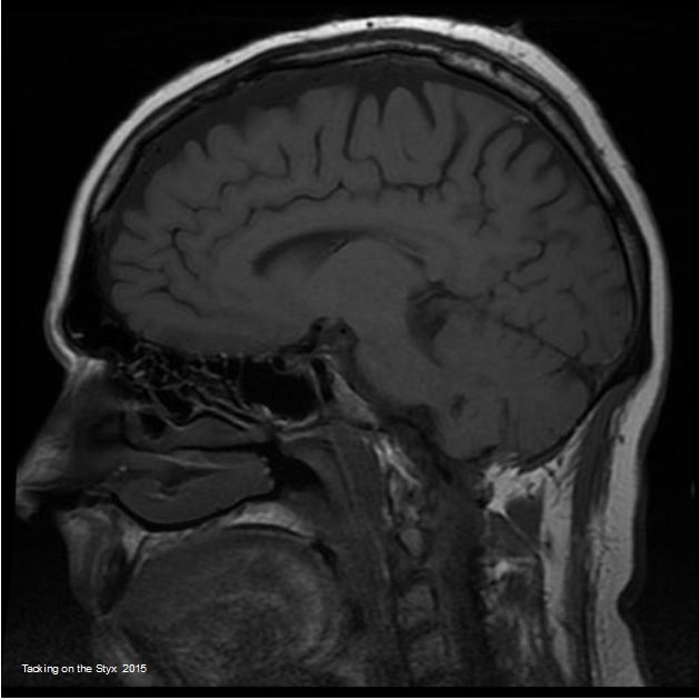
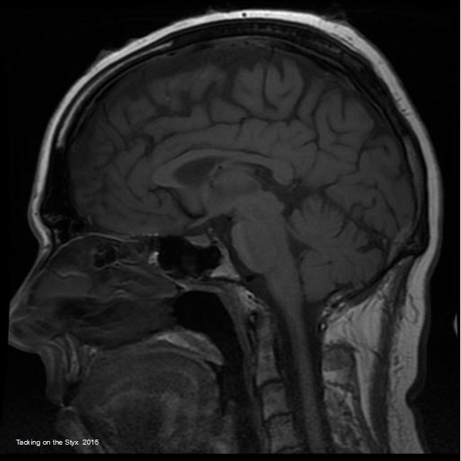
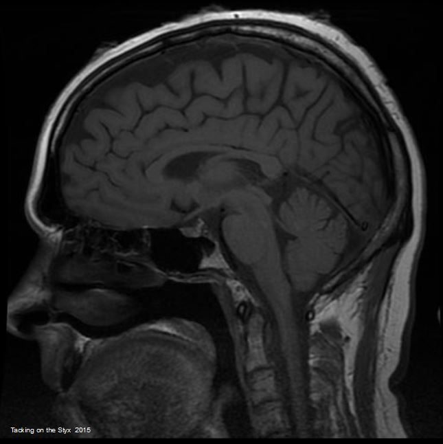
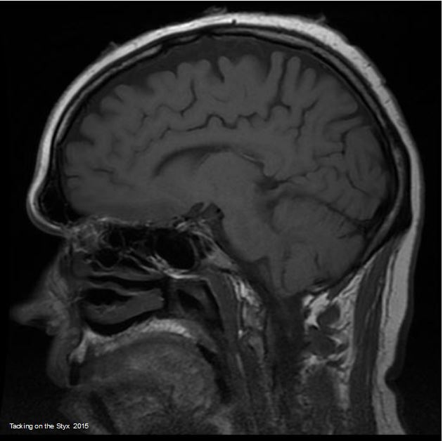
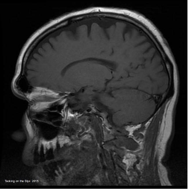
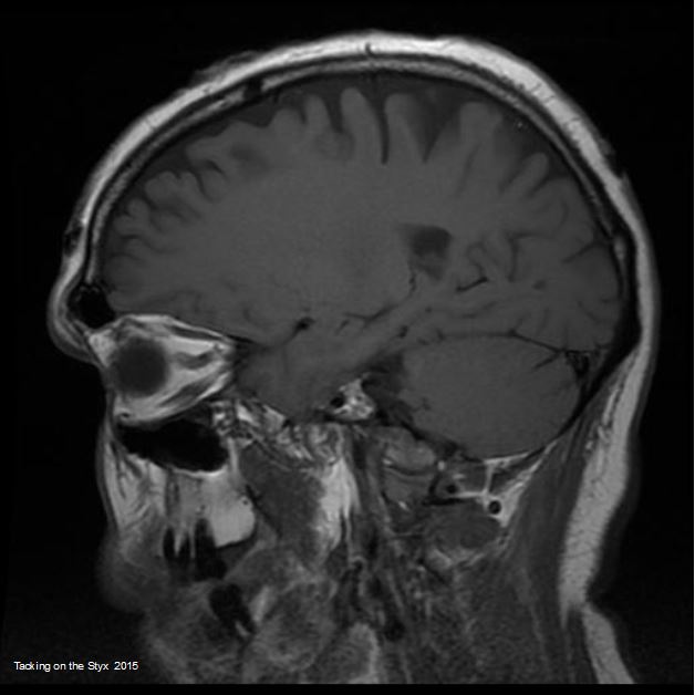


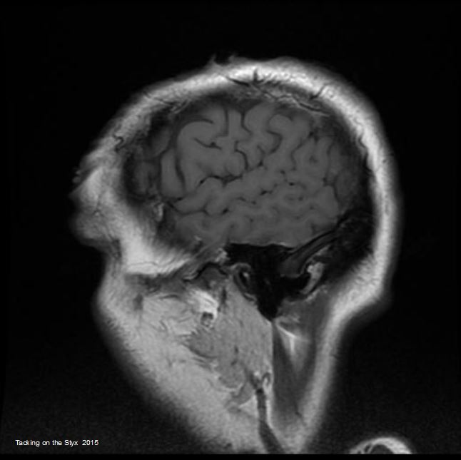
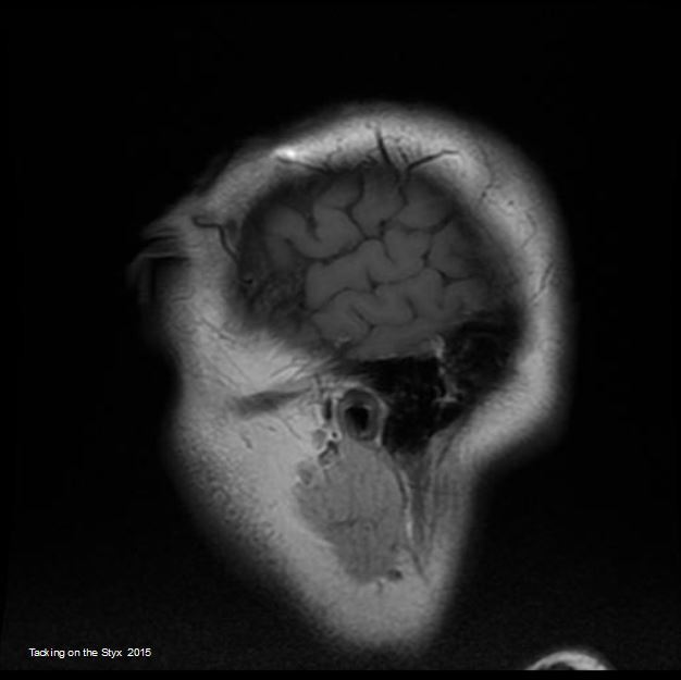

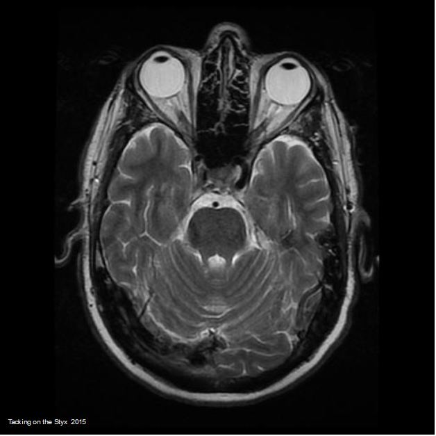


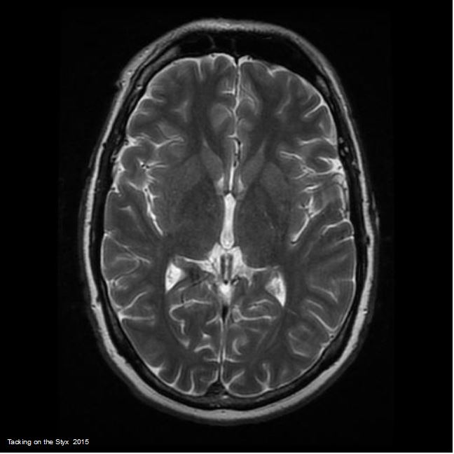


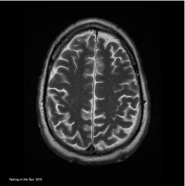

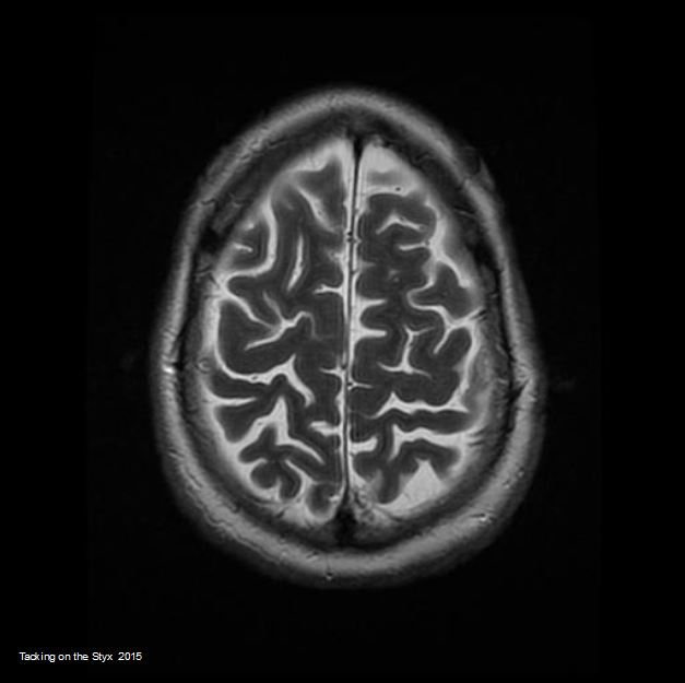
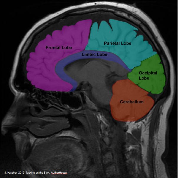
In addition to the lobes of the cerebrum, the medulla oblongata, and cerebellum, high quality can be found for the olfactory nerves, optic nerve, muscles of the eyes and neck, and many other features. Contact the author for additional imagery or go to Flicker sites here and here where you will find a larger number of images from the same MRI set.
The following may be used citing http://www.tackingonthestyx.com (Scroll further down for a gallery with several more coronal images)
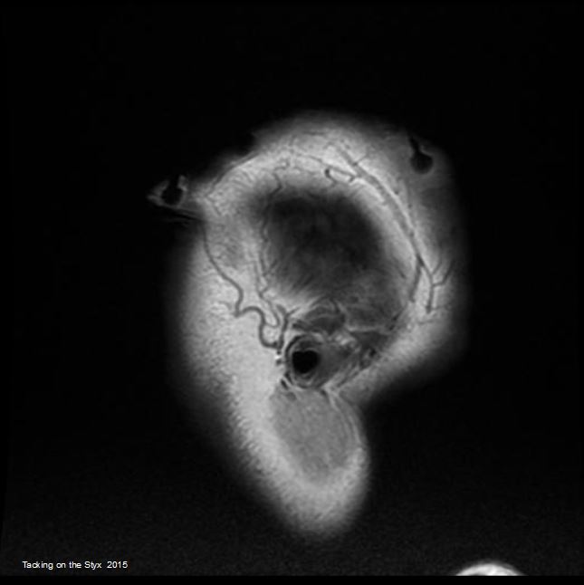
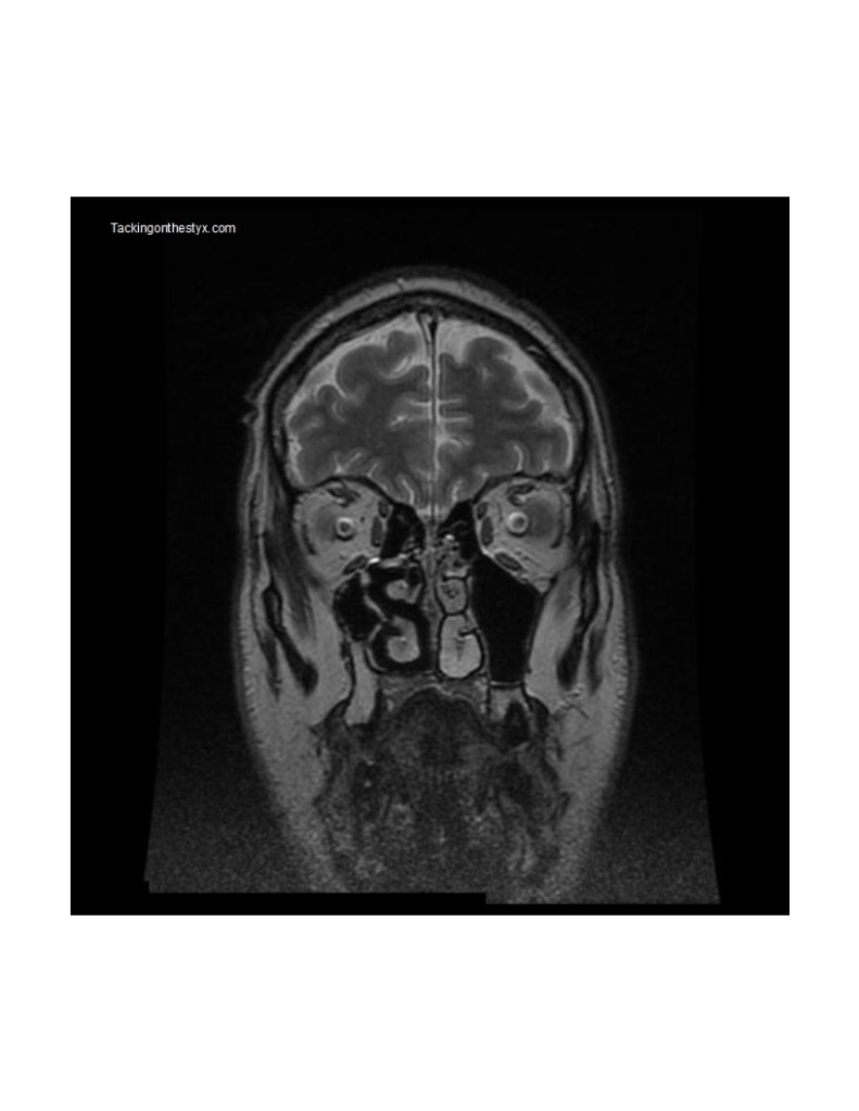
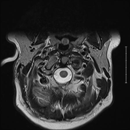
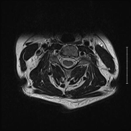
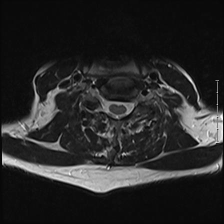
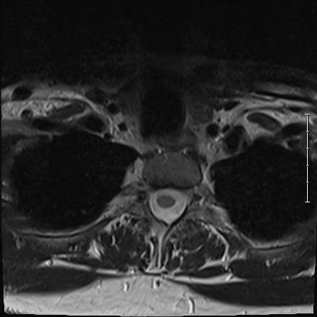
Coronal 2 Hatcher (only even numbers are used to correspond to scan slice uploaded). Features of note include olfactory bulb, medial rectus muscle, turbinate bones, orbital gyrus, olfactory sulcus.

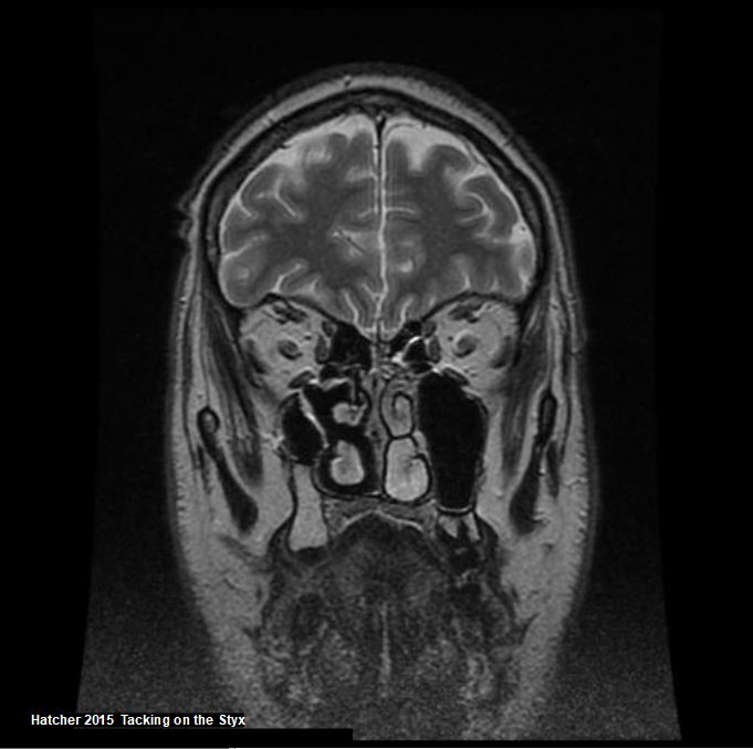
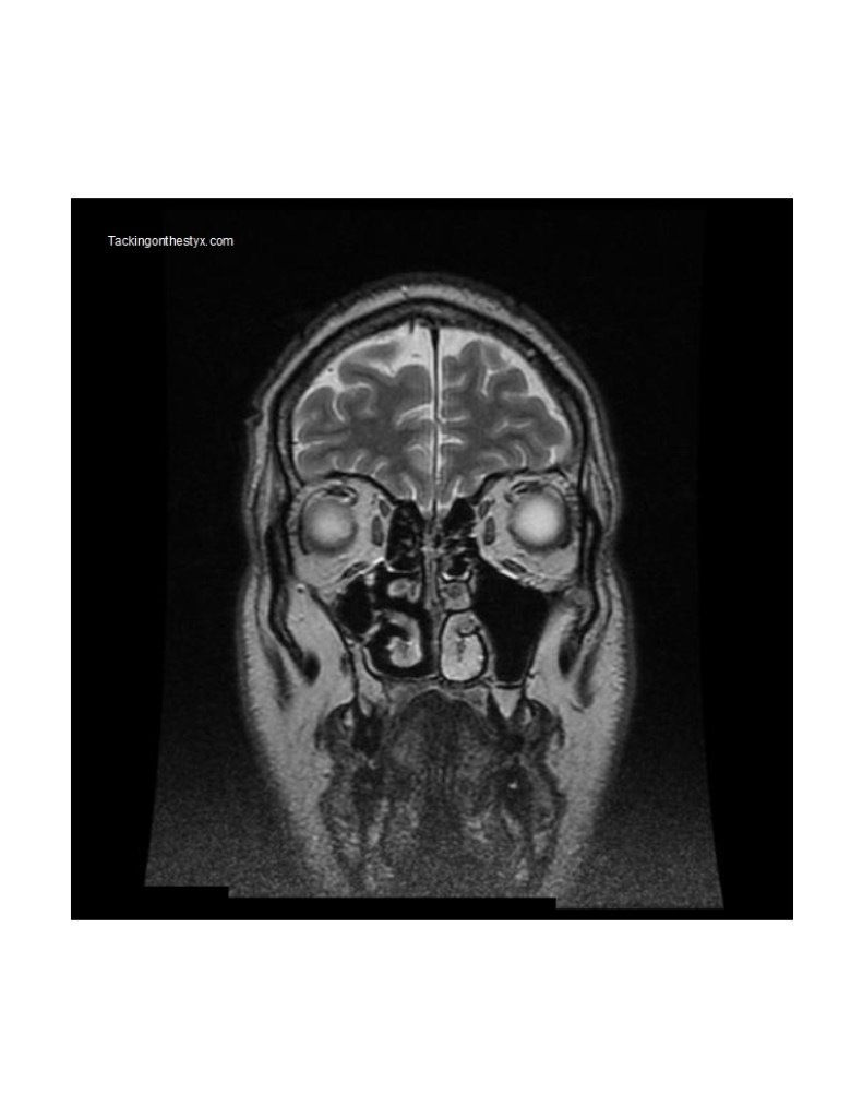
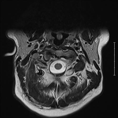
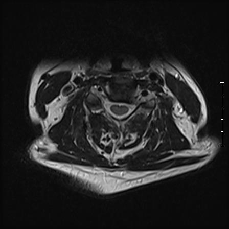
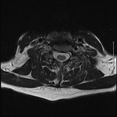
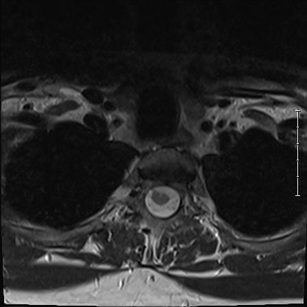
Coronal 4 Hatcher
Features of note include frontal gyrus, orbital gyrus, optic nerve, ethmoid sinus, maxillary antrum, turbinate bones.
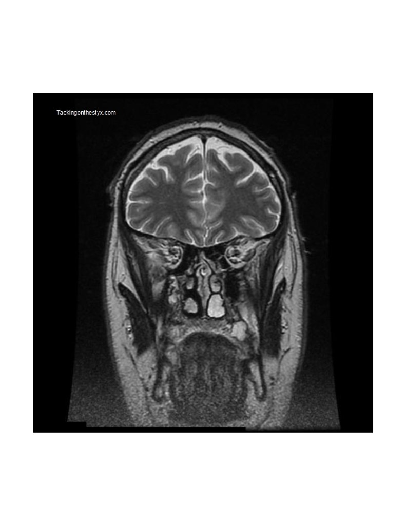
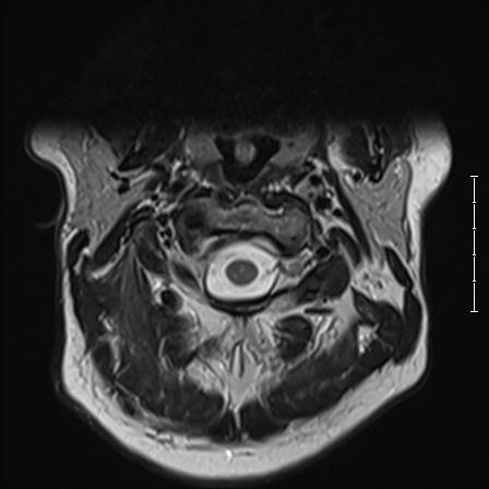
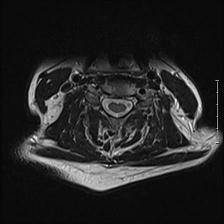
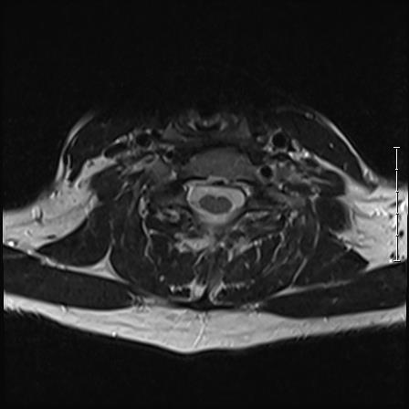
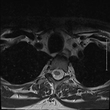
Coronal 6 Hatcher
Features of note include frontal lobes, white matter, grey matter, and turbinate bone.

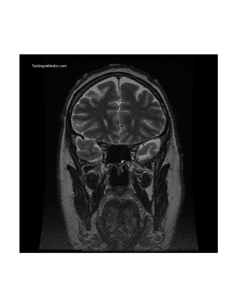
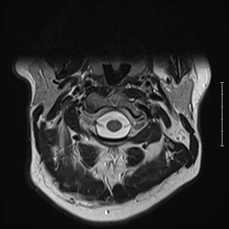
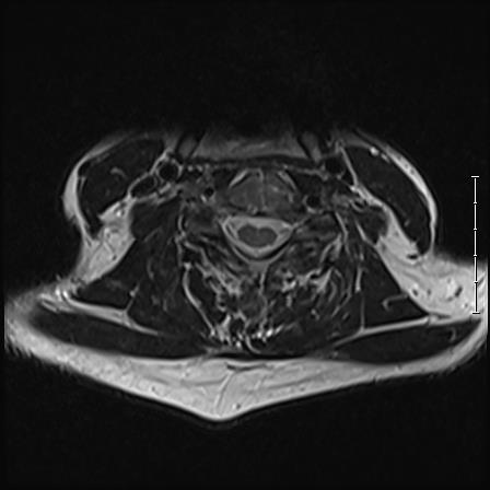
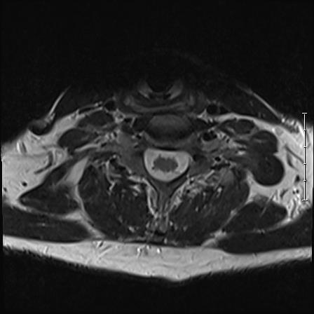
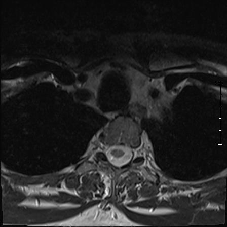
Coronal 8 Hatcher
Features of note include masseter muscle, temporalis muscle and frontal lobes.

Coronal 10 Hatcher
Features of note include superior sagittal sinus, sylvian fissure.

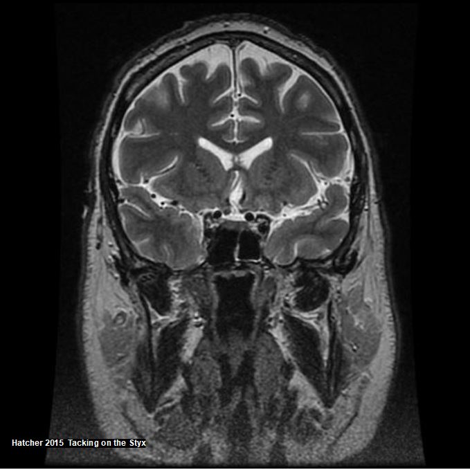
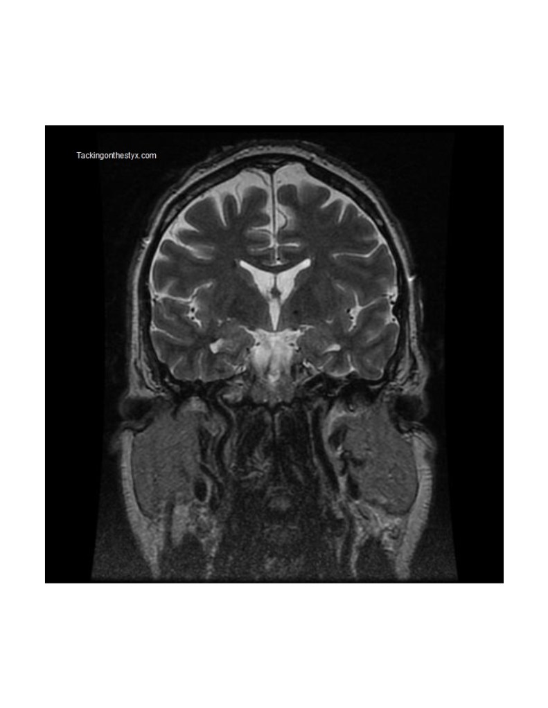
Coronal 12 Hatcher
Features of note include temporal lobes, frontal lobes, meso-temporal sclerosis, buccinator muscle, risorius muscle, anterior lateral ventricle.

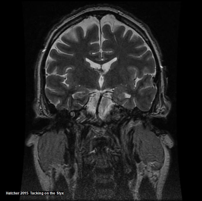
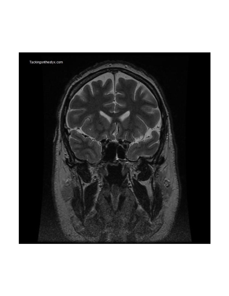
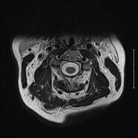
Coronal 14 Hatcher
Features of note include internal carotid arteries, parotid gland, nasopharynx, mandibular division.
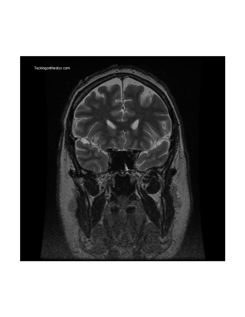
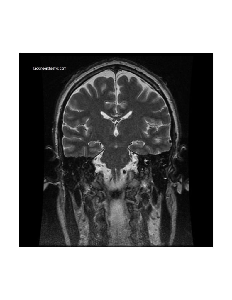
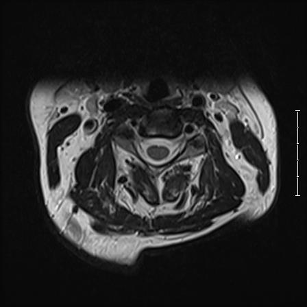
Coronal 16 Hatcher
Features of note include corpus callosum, interhemispheric fissure, sphenoid sinus, globus pallidus, internal carotid artery.

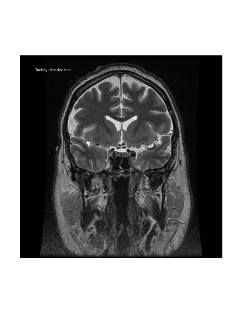
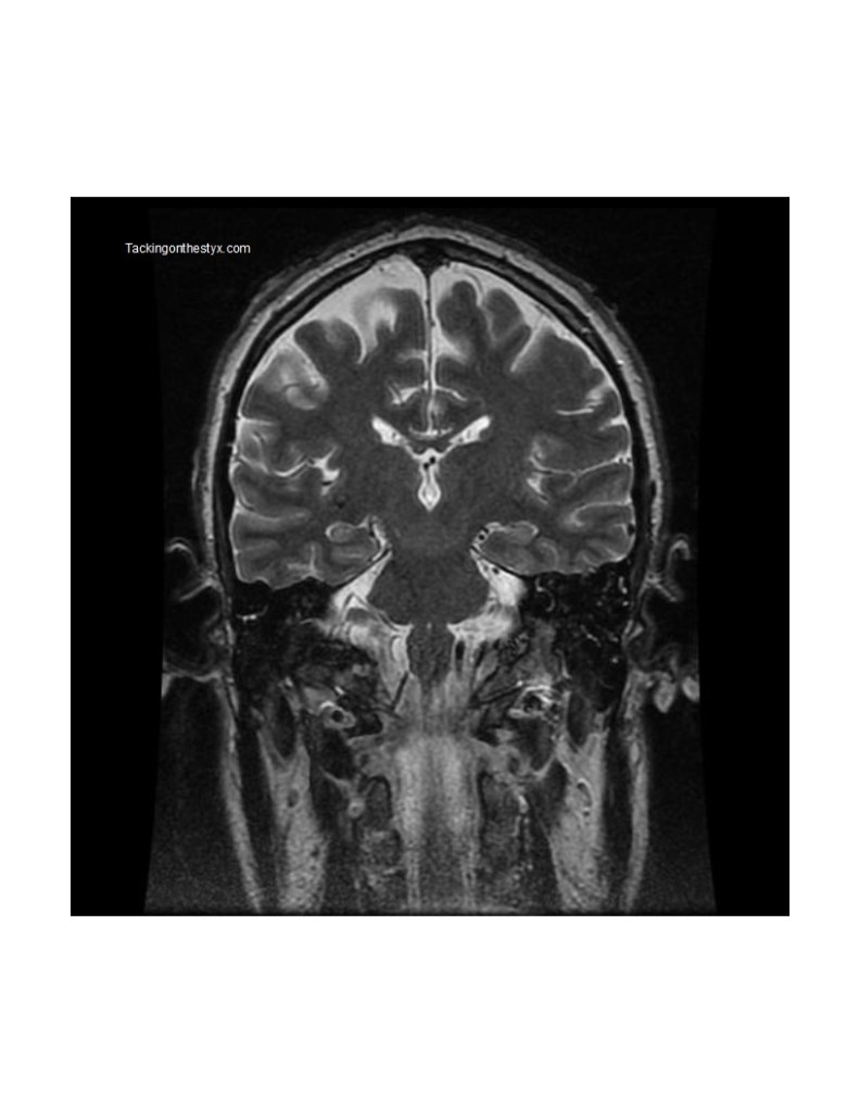
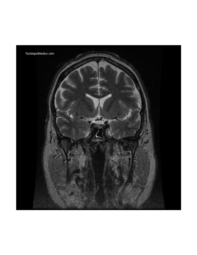
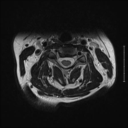
Coronal 18 Hatcher
Features of note include pituitary gland, optic tract, clivus, septum pellucidum, temporal lobe.

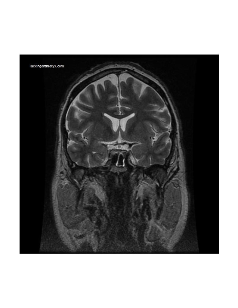
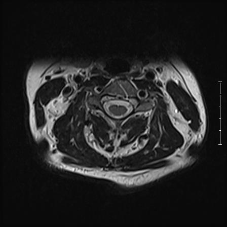
Coronal 20 Hatcher
Features of note include auditory canal, cochlea.
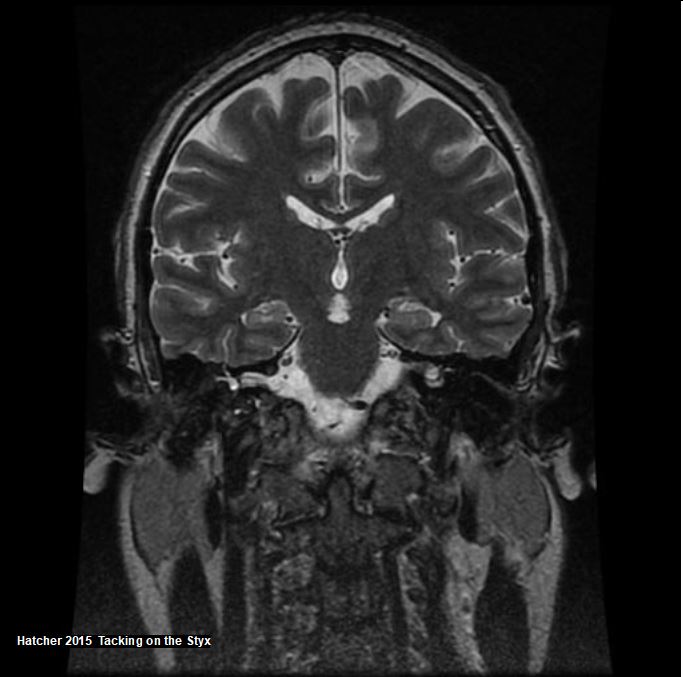
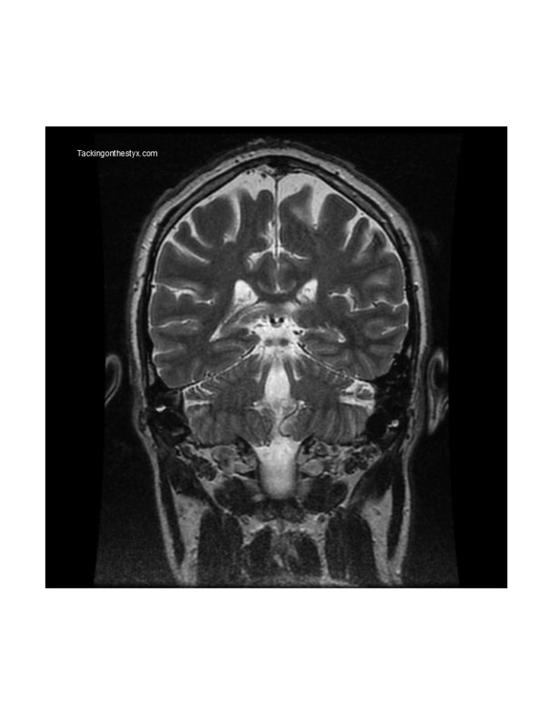
Coronal 22 Hatcher
Features of note include hippocampus, interpeduncular cistern, inner ear, pons, corpus callosum, superior sagittal sinus, preganglionic segment, third cranial nerve.
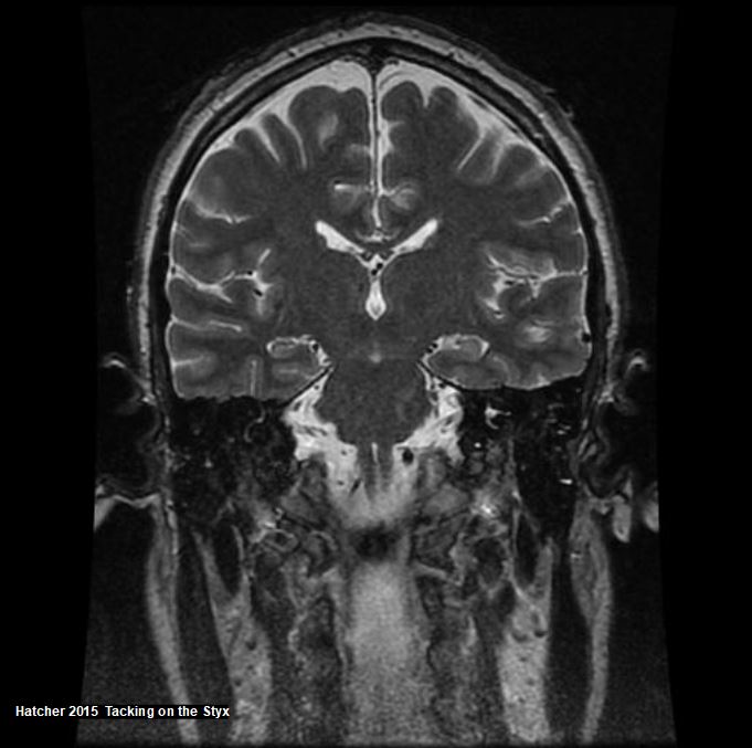
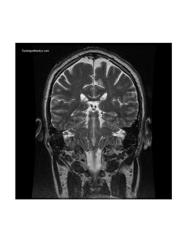
Coronal 24 Hatcher
Features of note include hippocampus, pons, thalamus, ambiens cistern, parahippocampal gyrus, temporal pole, cerebral peduncle.
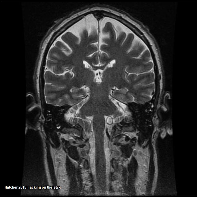
Coronal 26 Hatcher
Features of note include cerebellar peduncle, spinal cord, fornix, cerebral peduncle, medulla oblongata, brain stem, tentorium cerebelli.
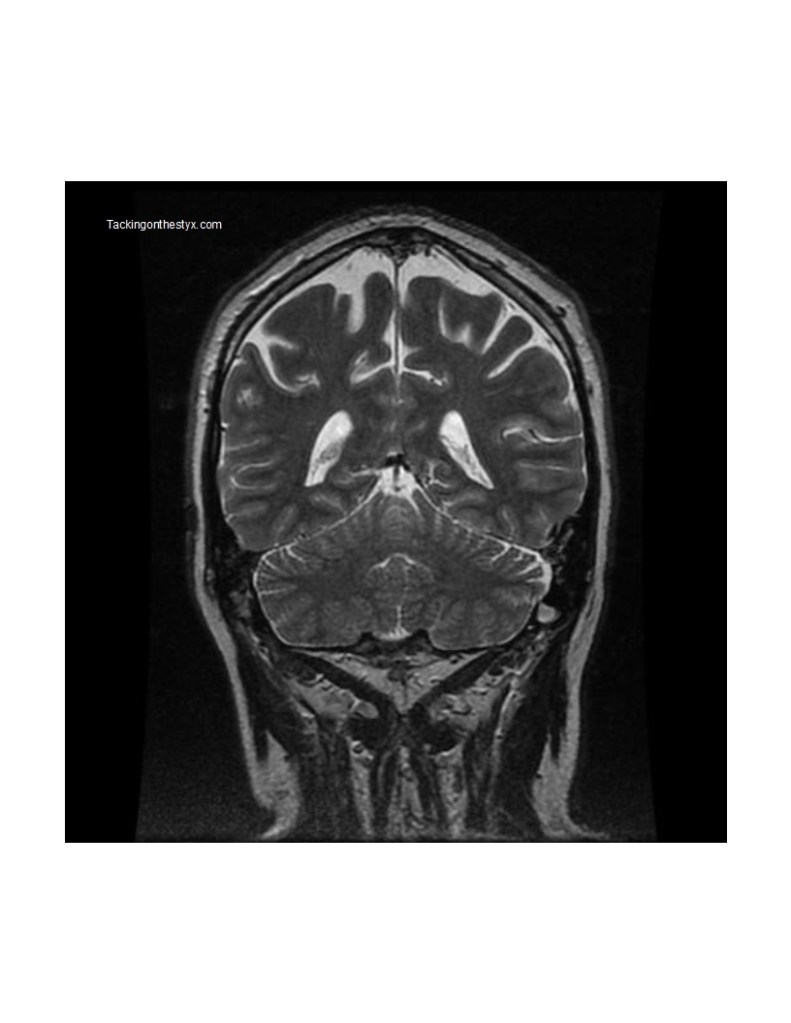
Coronal 28 Hatcher
Features of note include tentorium cerebelli, flocculus, thalamus, colliculus, fornix, cingulate sulcus, cingulate gyrus, parietal lobe.

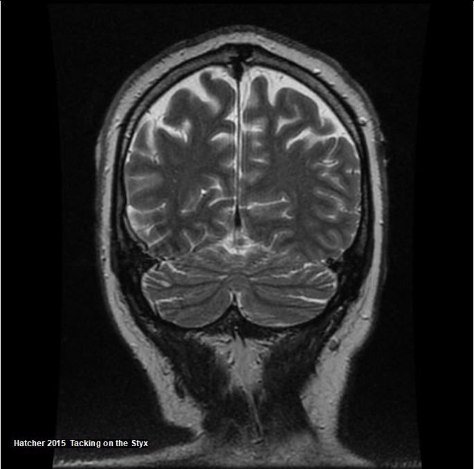
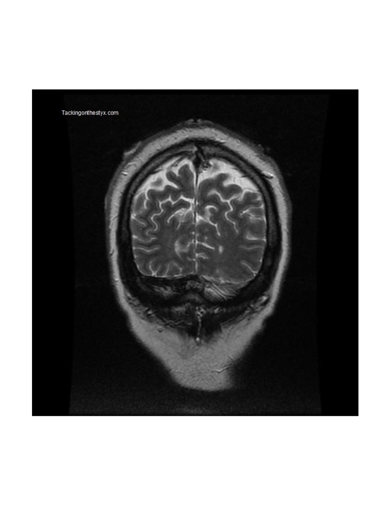
Coronal 30 Hatcher
Features of note include cerebellum, lateral ventricle.

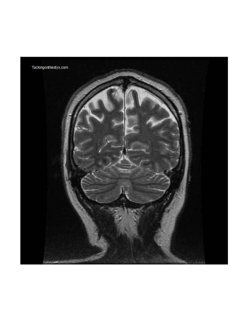
Coronal 32 Hatcher
Features of note include calcarine sulcus, cingulate sulcus, cerebellum.

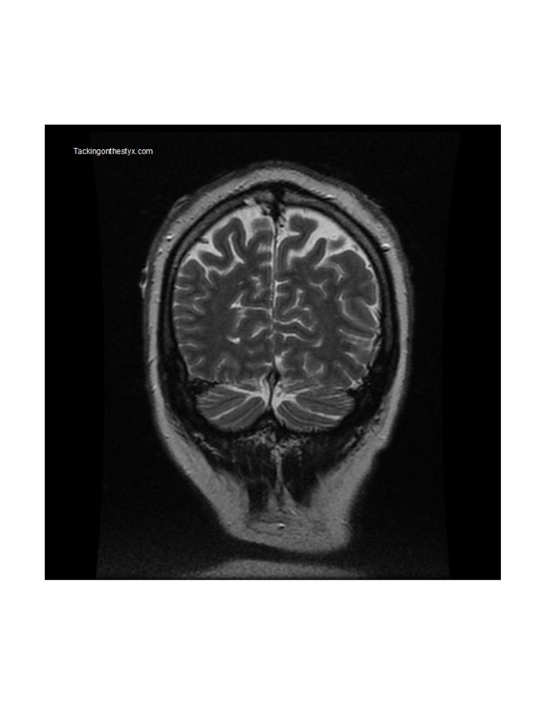
Coronal 34 Hatcher
Features of note include parietal lobe.

Coronal 36 Hatcher
Features of note include superior parietal lobule, inferior parietal lobule, intraparietal sulcus, parietal lobe, straight sinus.

Coronal 38 Hatcher
Features of note include intraparietal sulcus, precuneus, straight sinus.


Images below (with detailed condition diagnosis) may be used freely with citation to this website.
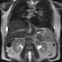
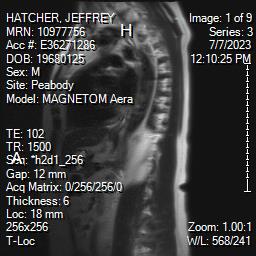
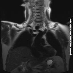
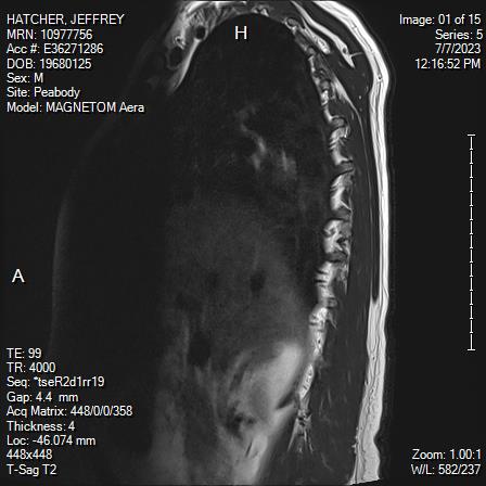
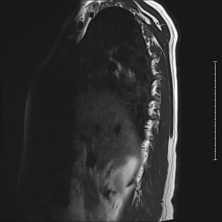
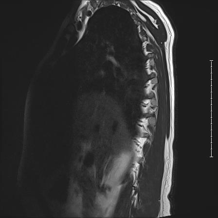
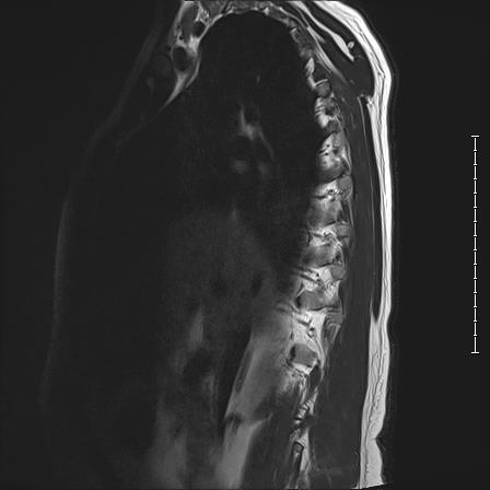
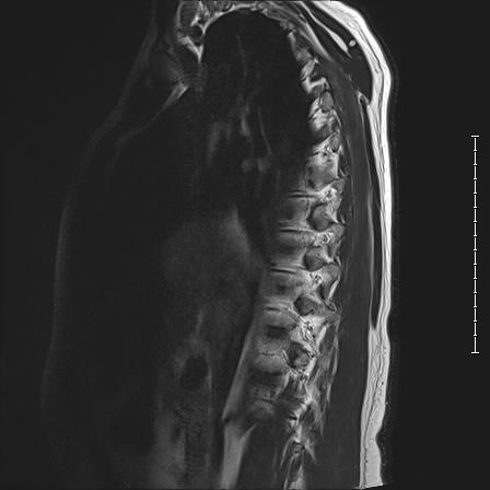
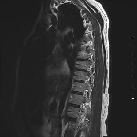
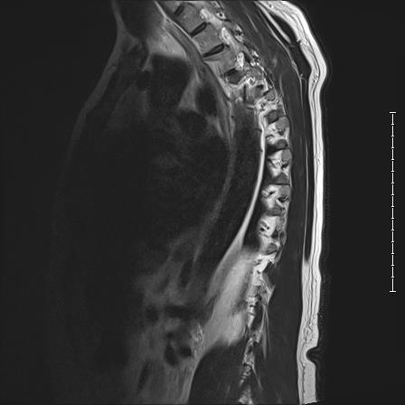
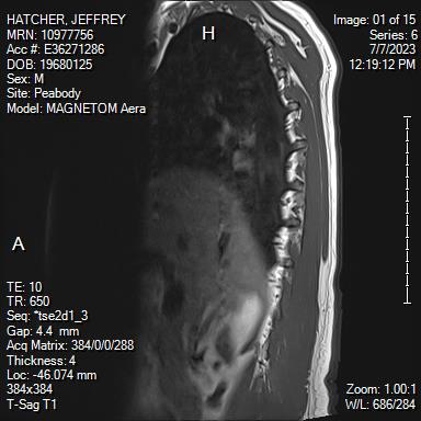
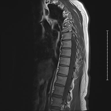
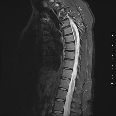
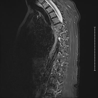
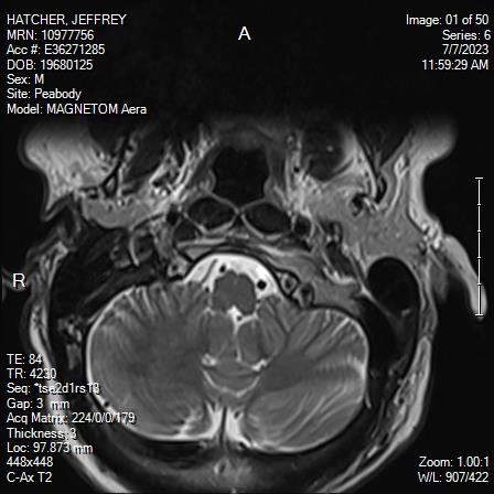
White Anglo-American male, 55 years of age with mild scoliosis and disk degeneration. MRI taken July, 2023 at Salem Hospital MRI facility using a MAGNETOM Aera model, 4 Centennial Drive, Peabody, Massachusetts.
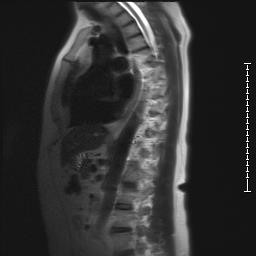
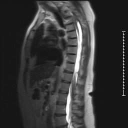
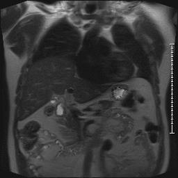
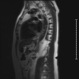
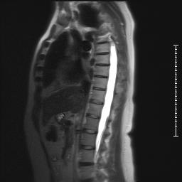
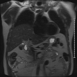
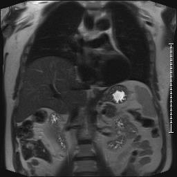
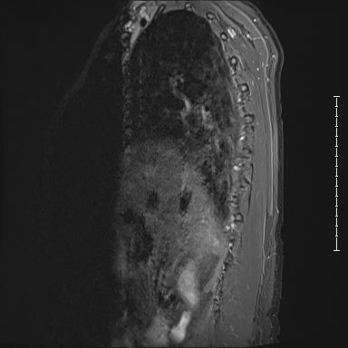
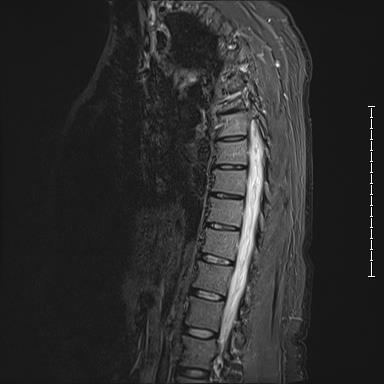
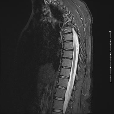
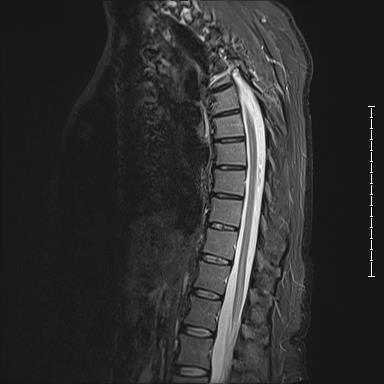
FORMAL REPORT ON THORACIC IMAGES:
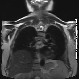
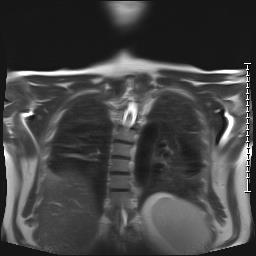
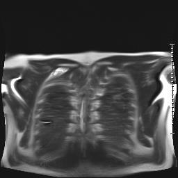
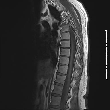
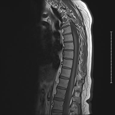
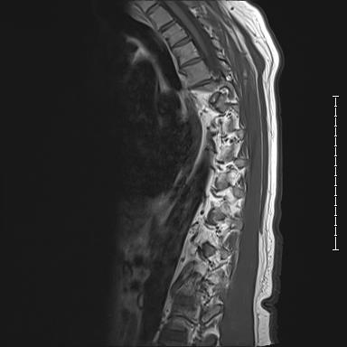
Mild to moderate scoliosis. Mild thoracic degenerative disc disease with mild disc desiccation and a few tiny posterior disc bulges, for example at T7-T8.
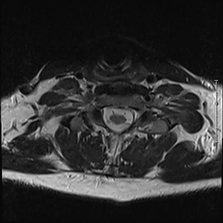
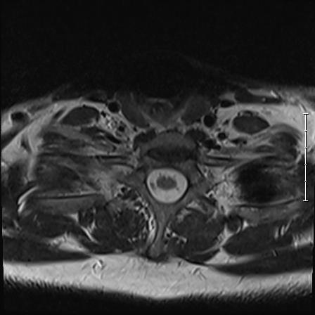
Thoracic MRI series 1


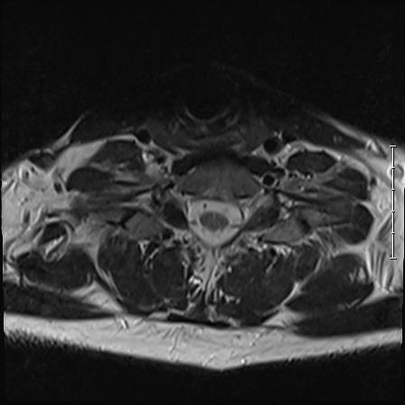
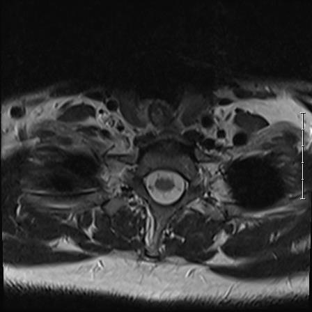
Thoracic MRI series 2


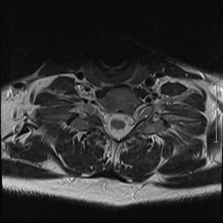
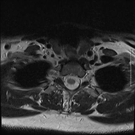
Thoracic MRI series 3


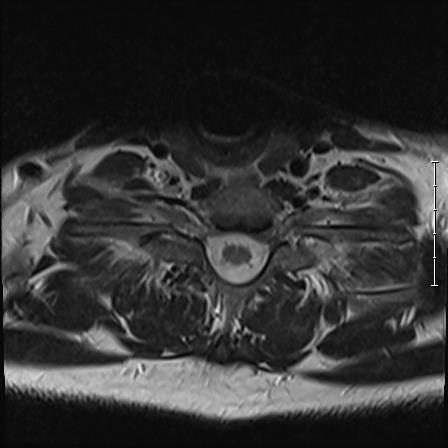
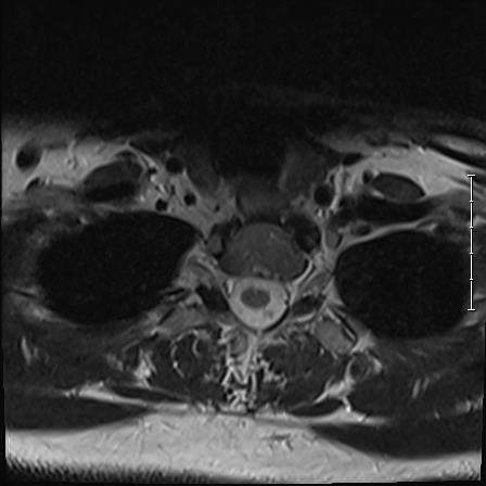
Thoracic MRI series 4


Thoracic MRI series 5


Thoracic MRI series 6


Thoracic MRI series 7


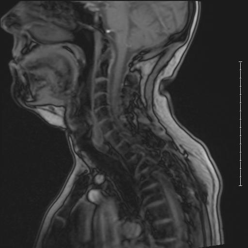
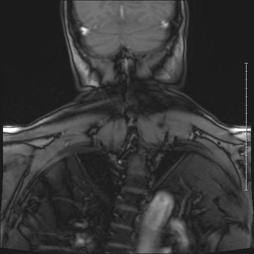
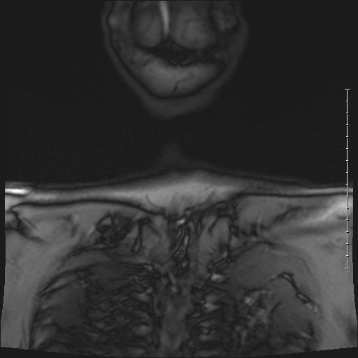
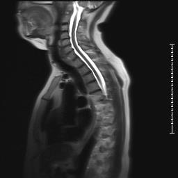
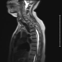
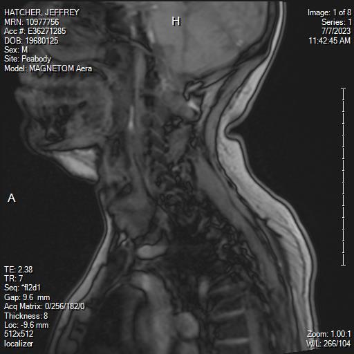
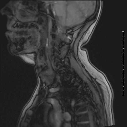
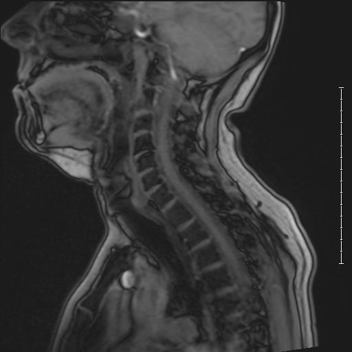
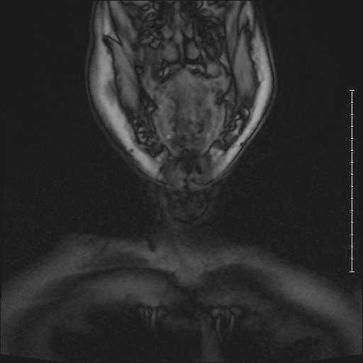
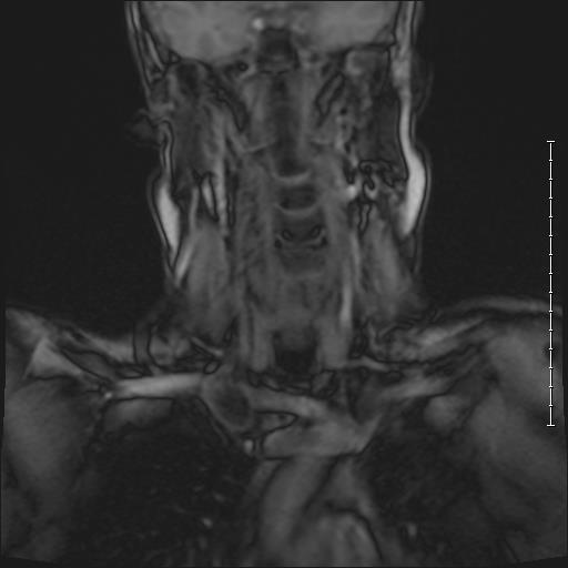
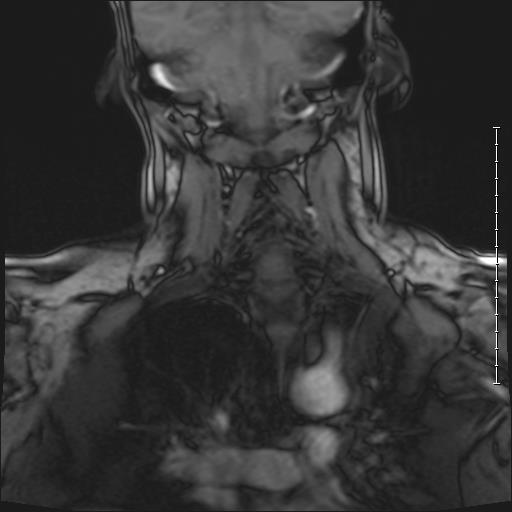
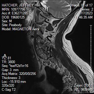
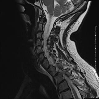
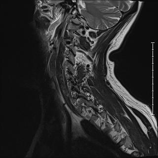
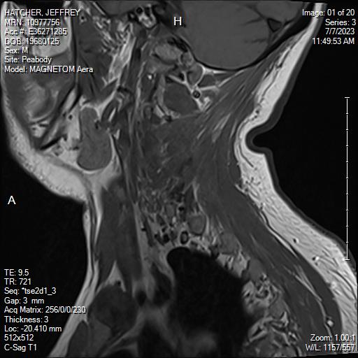
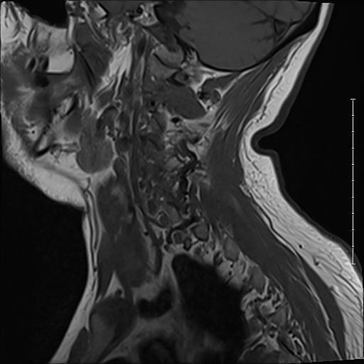
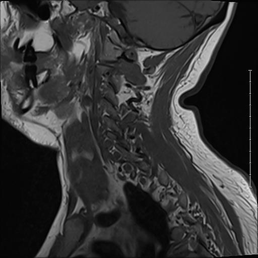
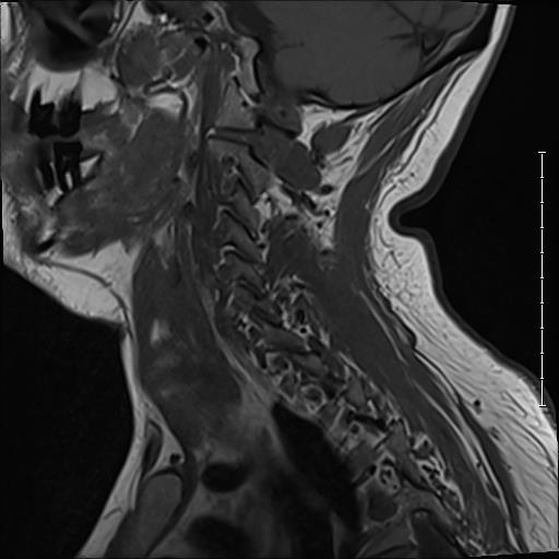
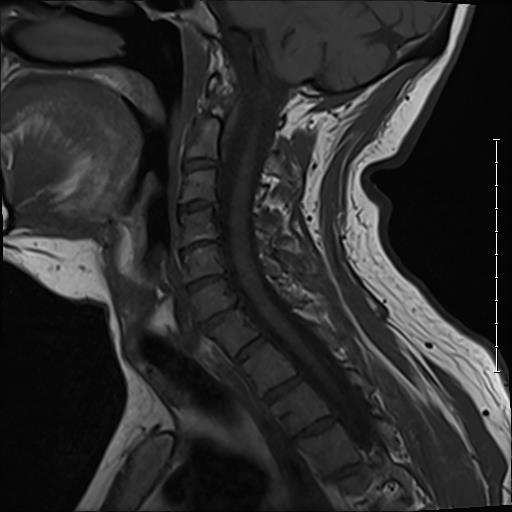
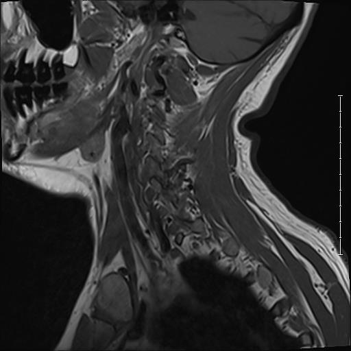
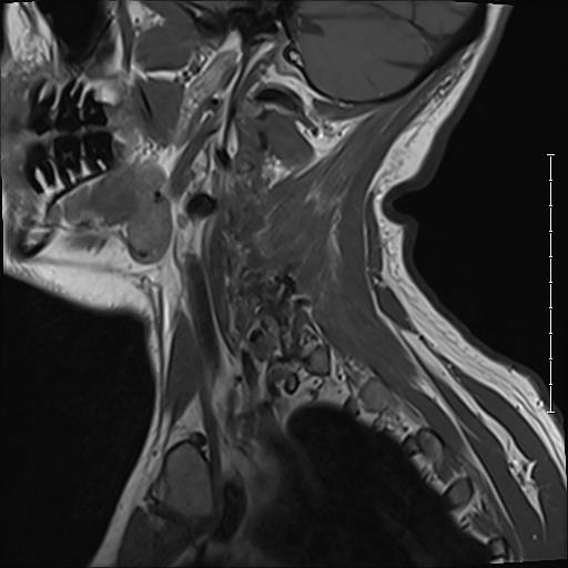
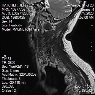
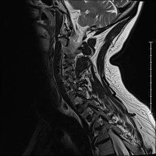
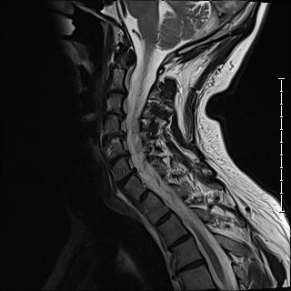
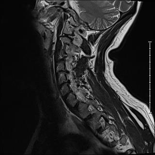
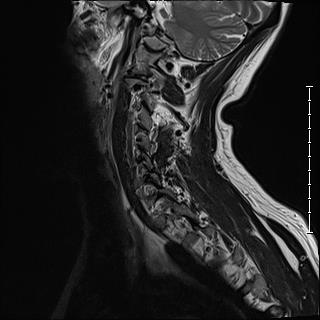
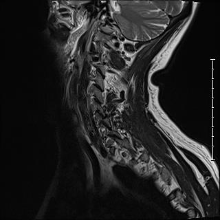
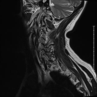
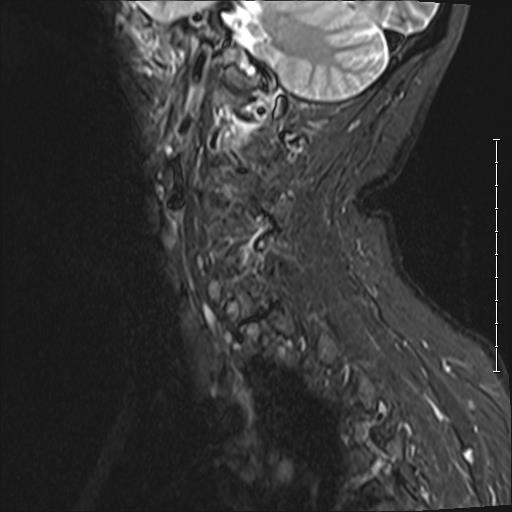
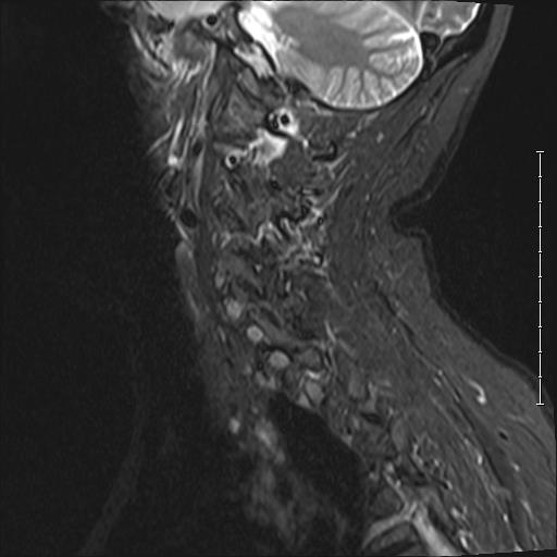
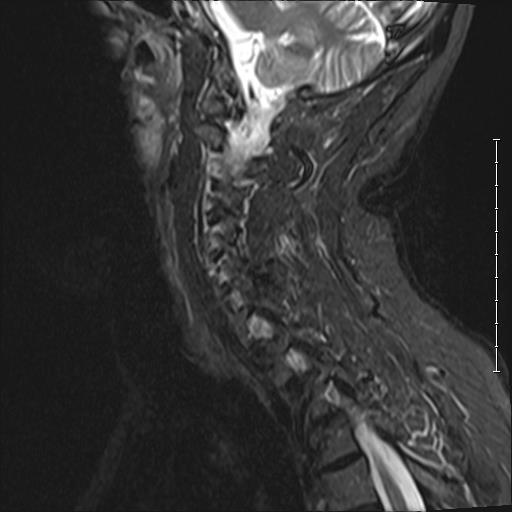
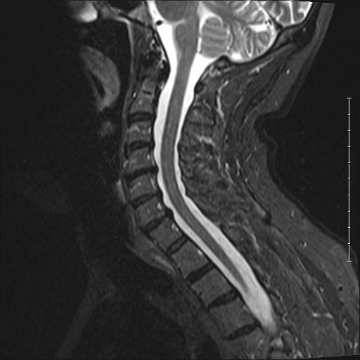
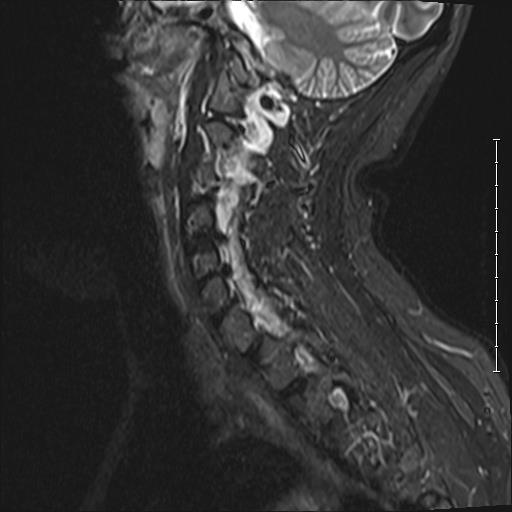
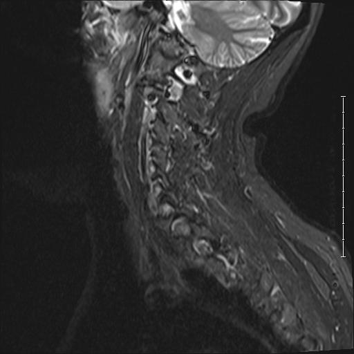
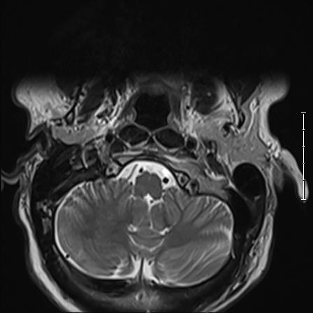
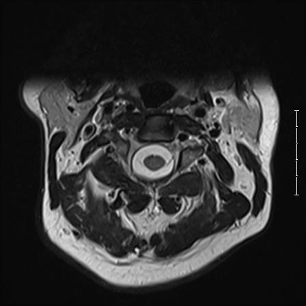
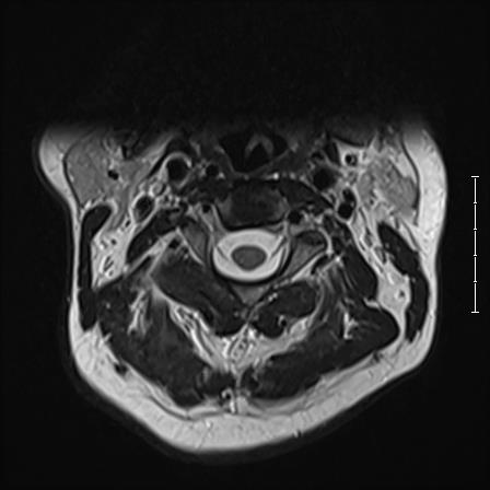
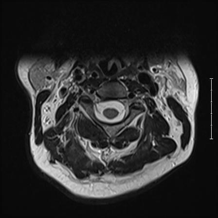
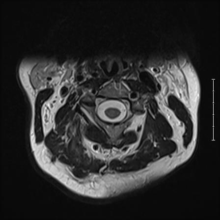
FORMAL REPORT ON CERVICAL IMAGES:
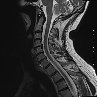
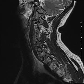
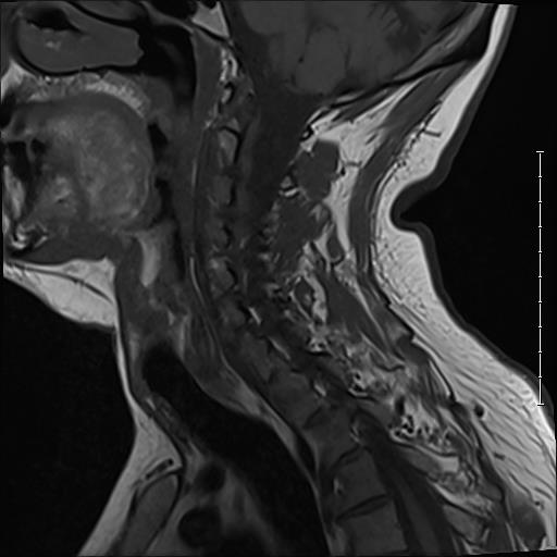
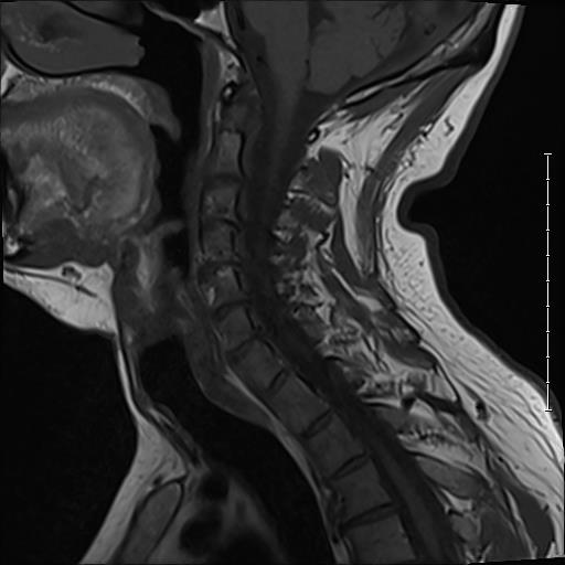
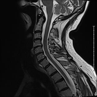
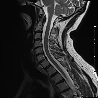
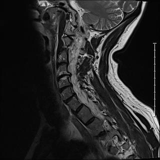
“The contents the posterior fossa are unremarkable. Cervical vertebra are preserved in height and AP alignment. There is at least mild scoliosis. Mild to moderate degenerative disc disease with disc desiccation at most levels. Small multilevel posterior disc osteophyte complexes, most notable from C3-4 through C6-7. Mild type I degenerative Modic endplate signal changes at C4-5. No evidence of acute compression fracture. Mild multilevel facet arthropathy. Mild effacement of ventral CSF particularly from C4-5 through C6-7. Mild multilevel facet arthropathy.”
Cervical MRI series 1


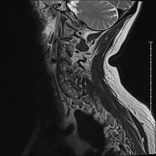
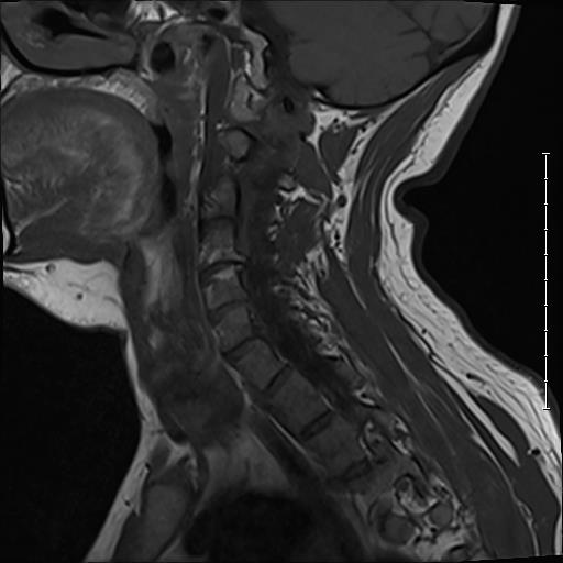
Cervical MRI series 2

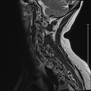


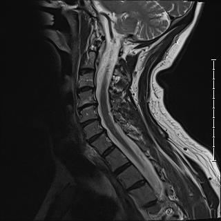


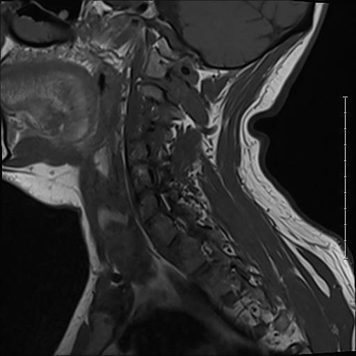
Cervical MRI series 3


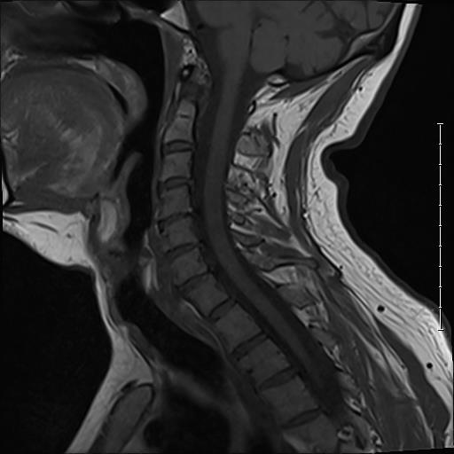

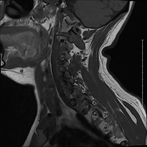


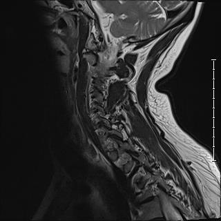
Cervical MRI series 4


Cervical MRI series 5


Cervical MRI series 6


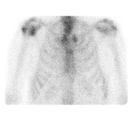
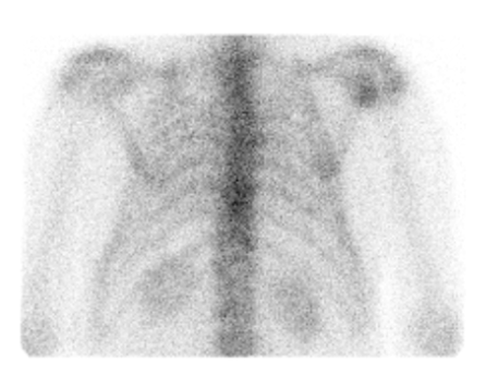
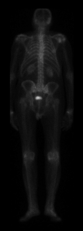
Thoracic and whole-body bone density scans
BDS of ninety-year-old male. Images taken from NovaPACS Diagnostic Viewer Study Browser v. 8.8. Lawrence General Hospital, Lawrence, MA on April, 2024. Formal report unavailable.

H.H. Smith, 1852. A system of operative surgery: based upon the practice of surgeons in the United States, and comprising a bibliographical index and historical record of many of their operations, during a period of two hundred years. Philadelphia: Lippincott, Grambo and Co.
